(UNDER CONSTRUCTION)

OUR VISION

The AURL pioneers innovative solutions in biomedical imaging and therapy by integrating multi-modal ultrasound with photoacoustic technologies. Our mission is to address significant challenges in healthcare by engineering cutting-edge hybrid imaging systems. These systems act as robust platforms for clinically translated theranostics that seamlessly fuse diagnostics and treatment. In particular, we are focused on developing technologies for disease monitoring and ultrasound stimulation to treat brain disorders.

AURL 은 다중 모드 초음파와 광음향 기술을 통합하여 의생명 영상 및 치료 분야의 혁신적인 솔루션을 개척합니다. 진단과 치료를 매끄럽게 융합하는 임상중개 테라노시스(theranostics)를 위한 강력한 플랫폼 역할을 하는 최첨단 하이브리드 영상 시스템을 개발하여 헬스케어의 중대한 과제들을 해결하는 것을 목표로 연구를 수행하고 있습니다. 특히, 뇌질환 치료를 위한 질병 모니터링, 초음파 자극기술 등을 연구 및 개발하고 있습니다.

BIOMEDICAL MULTI-MODAL ULTRASOUND IMAGING

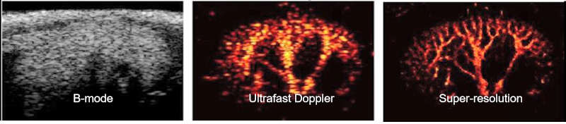
To provide a comprehensive understanding of complex diseases like cancer and cardiovascular conditions, we develop multi-modal ultrasound platforms. By integrating traditional B-mode sonography with advanced functional techniques—such as elastography (for tissue stiffness), thermal strain imaging (for composition), Doppler imaging (for hemodynamics), and contrast-enhanced imaging (for blood perfusion)—we can capture anatomical, mechanical, compositional, and optical information from a single imaging plane, simultaneously.


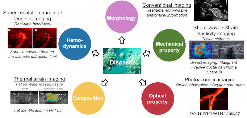
FIG. Multi-modal ultrasound imaging technologies

SUPER-RESOLUTION ULTRASOUND IMAGING

Standard ultrasound is limited in its ability to visualize microvasculature, which is critical for diagnosing diseases like chronic kidney disease and atherosclerosis. We are developing super-resolution ultrasound technologies that shatter the acoustic diffraction limit. By combining ultrafast imaging, advanced clutter filtering, and novel microbubble localization techniques, we can identify individual microvessels with unprecedented spatial resolution, revealing details up to five times finer than conventional methods.


FIG. Imaging on mouse kidney: B-mode, Ultrafast Doppler, Super-resolution imaging (x5 better resolution)

3-D ULTRASOUND IMAGING

To move beyond the limitations of 2D imaging, we are developing next-generation volumetric ultrasound systems. While traditional mechanical probes suffer from motion artifacts and slow speeds, we focus on electronic scanning with fully-sampled matrix arrays. This approach enables real-time, multi-planar visualization of tissue, essential for providing more accurate diagnoses, improving surgical guidance, and effectively monitoring therapy.


FIG. (a-c) Multi-planar imaging capabilities; real-time (d-e) C-scan and (f) Volumetric imaging

PHOTOACOUSTIC IMAGING

PAI is an emerging modality that uniquely combines the high contrast of optical imaging with the deep penetration of ultrasound. By detecting laser-induced ultrasound waves from the body's natural chromophores, our PAI systems provide real-time images of both anatomical structures and crucial physiological functions, such as oxygen saturation—a key indicator of cancer progression and metastasis.


FIG. Mouse brain imaging (Image courtesy of Chonnam University, Dr. Changho Lee), Enhanced light delivery scheme for Photoacoustic imaging (Featured on the cover of IEEE Transactions on UFFC, Aug, 2017)

NANOTECHNOLOGIES FOR MOLECULAR PHOTOACOUSTIC IMAGING

To enhance the power of PAI, we engineer novel nanotechnologies for molecular theranostics. Our work includes the development of optically-triggered phase-transition nanodroplets. These agents remain dormant as they accumulate in target tissues like tumors and, upon laser activation, vaporize into highly visible microbubbles. This process generates significantly stronger photoacoustic signals and creates a powerful dual-mode contrast agent for both photoacoustic and ultrasound imaging, enabling targeted diagnostics and therapy.


FIG. Perfluorocarbon-based phase-transition droplet for Photoacoustic imaging

Research Sponsors